The Age of Tomosynthesis Arrives
According to geometrictomography.com, tomography denotes an “area of mathematics dealing with the retrieval of information about a geometric object from data about its sections by lines or planes…” Most people are familiar with tomographic medical imaging as CT. However, the technique for section imaging known as tomosynthesis has a long history. Today, thanks to recent technological advances, it is rising to prominence due to lower cost, less radiation exposure and greater efficiency than CT—with increasingly high quality imaging results. The age of tomosynthesis is upon us.
1914
Polish Radiologist Karol Mayer describes the principles of tomography.
1921
Frenchman Andre Edmond Marie Bocage patents the principles of tomography, without creating a working model.
1921
Frenchmen Felix Portes and Maurice Chausse publish and patent similar forms of tomography without a working model.
1934
Dutchman Bernard Zeidses des Plantes describes a tomography device in a PhD thesis and builds the first operational system.
1934
German Gustave Grossmann creates and markets the first commercial tomography system through his company Siemens-Reiniger-Veifa. The Grossmann tomograph becomes the most widely used system by the late 1930s.
1950s
Several additional companies also build commercial tomography equipment.
1960s
Tomographic systems enjoy growing popularity for thoracic and abdominal imaging.
1960-1970
The growth of digital detectors lead to the next generation of tomographic devices, most prominently high quality but costly CT.
1972
D.G. Grant coins the term “tomosynthesis” in a groundbreaking paper that describes the method of tomosynthesis reconstruction . . . and true tomosynthesis is born.
1970-1980
A variety of tomosynthesis devices come to market.
Mid-1980s
Researchers create a range of deblurring algorithms to compensate for tomosynthesis’ inherent imaging artifacts.
Late-1990s
Flat panel digital detectors lead to the growth of digital tomosynthesis. They boost device performance, enabling significantly higher quality and faster imaging and sparking a new era in tomosynthesis.
2000-2010
Tomosynthesis imaging comes of age as a digital imaging technology and is applied in a variety of clinical imaging settings – breast and chest imaging most prominently.
Today
A growing number of sophisticated image acquisition techniques and reconstruction algorithms enhance tomosynthesis imaging information and expand its usefulness into increasing applications.
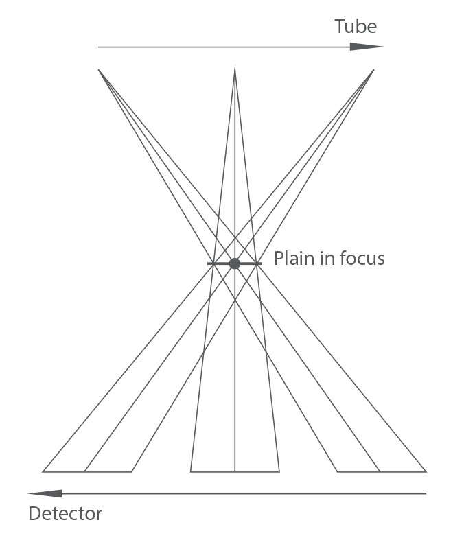
This is a generalized illustration of classical tomosynthesis. The x-ray tube and detector move in synchrony, one above and the other below the patient, as if around a central imaginary point. The layer where this point is set is the defined tomographic slice. The layers above and below are blurred out in the image acquired. The pivotal point may be varied in position and in this way repeat acquisitions of different tomographic slices may be obtained. These systems enjoyed a number of important applications in thoracic and abdominal imaging.

Digital tomosynthesis is a form of limited angle tomography that produces sectional, or slice, images from a series of projection images acquired as the x-ray tube moves over a prescribed path around the patient. The total angular range of movement is often less than 40°. Because the projection images are not acquired over a full 360° rotation about the patient, the resolution in the z direction (i.e., in the depth direction perpendicular to the x–y plane of the projection images) is limited. Therefore, tomosynthesis does not produce the isotropic spatial resolution achievable with computed tomography (CT). However, the resolution of images in the x–y plane of the reconstructed slices is often superior to CT, and its ease of use in conjunction with conventional radiography makes tomosynthesis a potentially very useful imaging modality.
